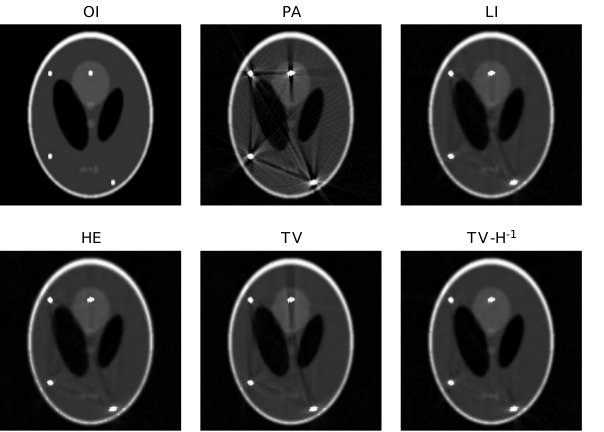
Medical images are often affected by noise or artifacts. A MOX research topic concerns the reduction of metal artifacts (MAR) in X-ray computerized tomography (CT), and the development of techniques and procedures for the pre-processing of medical images.
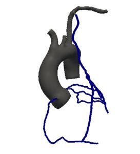
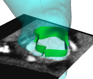
A second focus is the generation of automatic methods for the segmentation of different vessel district such as the carotid arteries, the aorta artery, the coronary tree and the heart chambers from different image modalities (e.g. CT scans, angio-CT scans, MRI images, etc…). To this aim some general purpose methods have been developed or adapted to ours applications. Moreover, specific methods have been studied and developed such as:
- automatic coronary tree generation from centerlines,
- plaque reconstruction in carotid arteries of patients with atherosclerosis,
- automatic segmentation of the aorta,
- semi-automatic left ventricle segmentation,
- parametric active contour segmentation methods,
- level set image segmentation through adaptive finite element methods,
- aortic valve leaflets reconstruction from CT images.
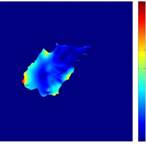
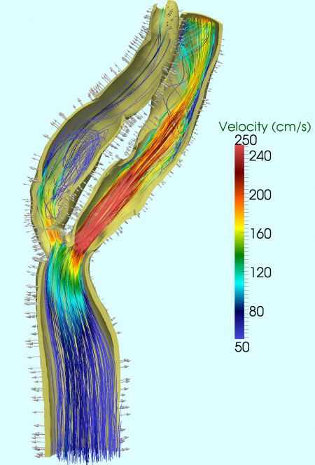
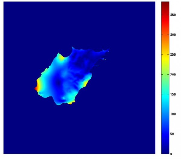
Another focus of this task is the development of methods for the analysis and the reconstruction of blood flow information from both phase contrast images (in which the pixel brightness is proportional to the velocity of the material) and from eco doppler signals. Velocity field are extremely useful in order to impose patient-specific boundary conditions, to perform validation of numerical simulations and in a data assimilation framework.
In addition to anatomical and velocity informations, also the knowledge of the three dimensional (3D) motion field of the arteries such as the carotid bifurcation is important for several applications, such as vessel compliance estimation, data assimilation or fluid-structure interaction validation. A widespread non-invasive method for imaging wall motion in clinical settings is 2D-cine-MRI, whereby a set of individual slices are acquired along the carotid bifurcation in which carotid wall motion is recorded. Within the Image Data Processing task , we proposed a new method to reconstruct the complete 3D carotid motion field starting from the 2D-cine-MRI slices and a static 3D-MRI data.
Publications:
- E. Faggiano, T. Lorenzi, A. Quarteroni, Metal artefact reduction in computed tomography images by a fourth-order total variation flow, Computer Methods in Biomechanics and Biomedical Engineering: Imaging & Visualization (2014), DOI: 10.1080/21681163.2014.94062
- E. Faggiano, L. Antiga, Interpolation-based reconstruction of human carotid dynamics from magnetic resonance images , Proceedings of SIMAI 2012 Biannual Congress, MSP – Computational and statistical methods for biomedical applications , Torino, 25-28 June 2012
- J. Bonnemain, E. Faggiano, S. Deparis, A. Quarteroni, and L. von Segesser. Segmentation and grid generation for numerical simulations of vad connections with patient-specific data. INTERNATIONAL JOURNAL OF ARTIFICIAL ORGANS Volume: 34 Issue: 8 Special Issue: SI Pages: 671-671, 2011.
- M. Sangalli, P. Secchi, S. Vantini, A. Veneziani, Efficient estimation of 3-dimensional centerlines of inner carotid arteries and their curvature functions by free knot regression splines, Journal of the Royal Statistical Society Ser. C, Applied Statistics, 58(3), pp. 285-306 (2009)
- R. Ponzini, C. Vergara, A. Redaelli, A. Veneziani, Reliable CFD-based estimation of flow rate in haemodynamics measures, Ultrasound in Medicine & Biology, 32(10), pp. 1545-1555 (2006)
- G. Abdoulaev, S. Cadeddu, G. Delussu, M. Donizelli, C. Manzi, L. Formaggia, A. Giachetti, E. Gobbetti, A. Leone, P. Pili, A. Schenine, M. Tuveri, A. Varone, A. Veneziani, G. Zanetti, A. Zorcolo, ViVA: The Virtual Vascular Project, IEEE Transactions on Information Technology in Medicine, 4(2), pp. 268-274 (1998)
Thesis:
- M. Fedele – A Patient-Specific Aortic Valve Model based on Moving Resistive Immersed Surfaces , MSc in Mathematical Engeneering, Politecnico di Milano – Advisors: A. Quarteroni, E. Faggiano A. Laadhari – A.Y. ’12-’13
- R. Ferrero – Numerical study of the fluid-dynamics in patient-specific coronary artery bypass grafts – MSc in Mathematical Engeneering, Politecnico di Torino – Advisors: L. Preziosi, A. Quarteroni, E. Faggiano – A.Y. ’12-’13
- F. Pizzi – Un approccio probabilistico per la segmentazione di immagini mediche tramite contorni attivi parametrici – MSc in Mathematical Engeneering, Politecnico di Milano – Advisors: A. Guglielmi, E. Faggiano – A.Y. ’12-’13
- M. Haile – Restauro di immagini tomografiche mediche tramite l’uso di metodi di image inpainting – MSc in Mathematical Engeneering, Politecnico di Milano – Advisors: S. Perotto, E. Faggiano – A.Y. ’12-’13
- N. Papucci – Adattazione anisotropa di griglia applicata alla segmentazione di immagini, MSc in Mathematical Engeneering, Politecnico di Milano – Advisor: S. Perotto – A.Y. ’11-’12
- E. Maculan – Simulazioni fluidodinamiche in vasi deformabili con moto della parete ricostruito da immagini mediche, MSc in Mathematical Engeneering, Politecnico di Milano – Advisor: A. Veneziani – A.Y. ’06-’07
- L. Cattaneo and C. Colciago – Analisi della diffusività di materiali porosi a partire da immagini – MSc in Mathematical Engeneering, Politecnico di Milano – Advisor: P. Zunino – A.Y. ’06-’07


