A new MOX Report entitled “An integrated heart-torso electromechanical model for the simulation of electrophysiological outputs accounting for myocardial deformation” by Zappon, E.; Salvador, M.; Piersanti, R.; Regazzoni, F.; Dede’, L.; Quarteroni, A. has appeared in the MOX Report Collection.
Check it out here: https://www.mate.polimi.it/biblioteca/add/qmox/14-2024.pdf
Abstract:
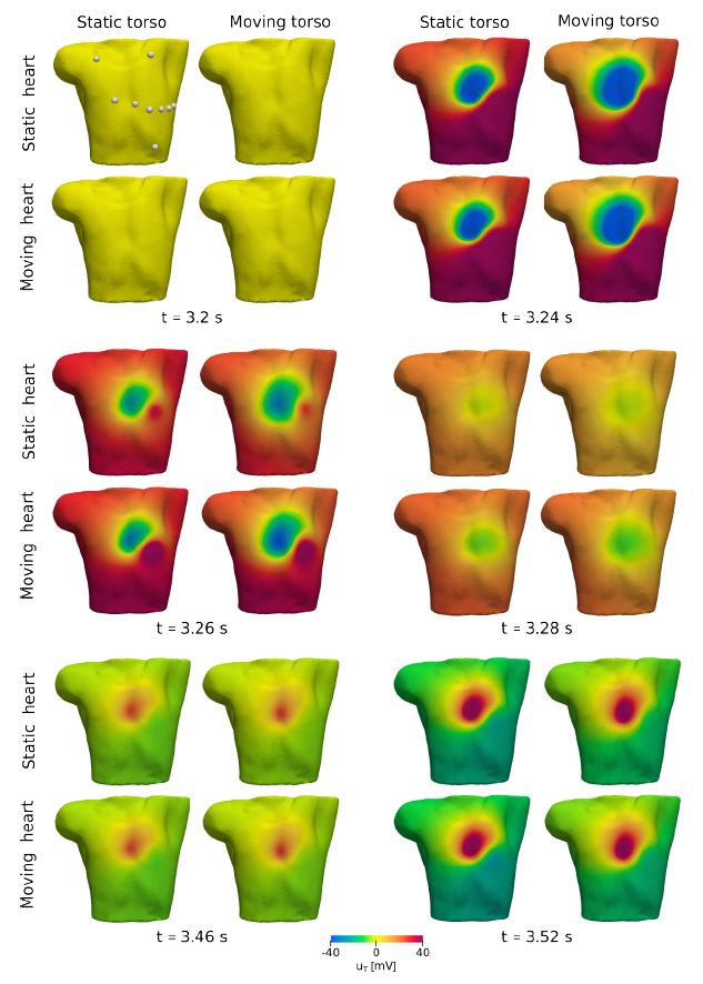
When generating in-silico clinical electrophysiological outputs, such as electrocardiograms (ECGs) and body surface potential maps (BSPMs), mathematical models have relied on single physics, i.e. of the cardiac electrophysiology (EP), neglecting the role of the heart motion. Since the heart is the most powerful source of electrical activity in the human body, its motion dynamically shifts the position of the principal electrical sources in the torso, influencing electrical potential distribution and potentially altering the EP outputs. In this work, we propose a computational model for the simulation of ECGs and BSPMs by coupling a cardiac electromechanical model with a model that simulates the propagation of the EP signal in the torso, thanks to a flexible numerical approach, that simulates the torso domain deformation induced by the myocardial displacement. Our model accounts for the major mechano-electrical feedbacks, along with unidirectional displacement and potential couplings from the heart to the surrounding body. For the numerical discretization, we employ a versatile intergrid transfer operator that allows for the use of different Finite Element spaces to be used in the cardiac and torso domains. Our numerical results are obtained on a realistic 3D biventricular-torso geometry, and cover both cases of sinus rhythm and ventricular tachycardia (VT), solving both the electromechanical-torso model in dynamical domains, and the classical electrophysiology-torso model in static domains. By comparing standard 12-lead ECG and BSPMs, we highlight the non-negligible effects of the myocardial contraction on the EP-outputs, especially in pathological conditions, such as the VT.


